
Technology behind the scene
Abstract:
Therapeutic ultrasound effects on living tissue might be grouped by two major categories: Thermal and Non-thermal. Non-focused ultrasound is capable of producing thermal therapeutic effects. Recent in vivo studies show that dissipated ultrasound beam is could increase the tissue temperature at a rate of 0.16°C/min (1 W/cm2, 1 MHz), up to 42°C. 3-MHz frequency increased tissue temperature at a faster rate than the 1-MHz frequency (Ashton, Chan). However, as the temperature increases, the normal blood flow to the area dissipates the heat, thus limiting further temperature rise. Focused ultrasound or high intensity focused ultrasound – HIFU is capable of reaching 70°C and beyond, where tissue destruction is inevitable. SonNext technology is solely based on Non-thermal effect of the therapeutic ultrasound, taking it several steps further to prevent any heat related issues.
Non-Thermal Ultrasound Distinct Features:
Frequency resonance hypothesis:
Acoustic Streaming.
Therapeutic ultrasound produces a combination of an acoustic streaming and cavitation effects that are difficult to isolate. Acoustic streaming is defined as the physical forces of the sound waves, generated by the areas of the high and low pressure, that provides a driving force capable of displacing ions and small molecules. At the cellular level, organelles and molecules of different molecular weight exist. While many of these structures are stationary, many are free floating and may be driven to move around more stationary structures. This mechanical pressure applied by the wave produces unidirectional movement of fluid along and around cell membranes.
Cavitation
Cavitation is defined as the physical forces of the sound waves on microenvironmental gases within fluid. As the sound waves propagate through the medium, the characteristic compression and rarefaction causes microscopic gas bubbles in the tissue fluid to contract and expand. It is generally thought that the rapid changes in pressure (caused by the leading and lagging edges of the sound wave), both in and around the cell, may cause damage to the cell. Substantial injury to the cell can occur when microscopic gas bubbles expand and then collapse rapidly, causing a “micro-explosion.” Although true micro-explosions, referred to as unstable cavitation, are not thought to commonly occur at therapeutic levels of ultrasound. Early studies investigating the gross effects of acoustic streaming and cavitation on cells showed growth retardation of cells in vitro, increases in protein synthesis, and membrane alterations. Combined, these results may suggest that ultrasound first “injures” the cell, resulting in growth retardation, and then initiates a cellular recovery response characterized by an increase in protein production.
Shear Stress
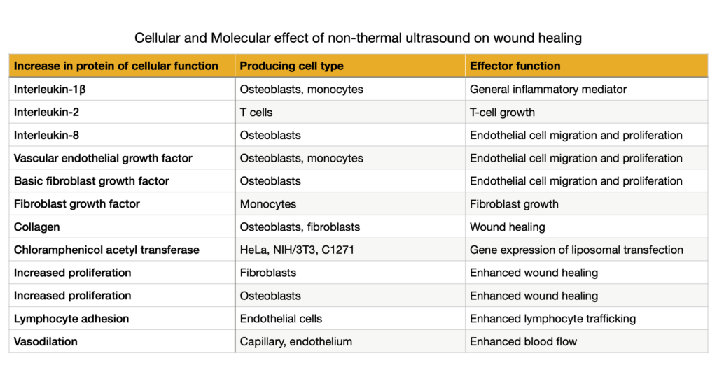
Shear stress of the ultrasound alters cellular membrane properties (cellular adhesion, membrane permeability, calcium flux, and proliferation), possibly activating signal-transduction pathways that lead to gene regulation Importantly, exposure to ultrasound caused an increase in intracellular calcium in fibroblasts, suggesting that the mechanical effects disrupt the normal function of the membrane, permitting leaking of calcium into the cell. After ultrasound exposure, the cells rapidly expelled the calcium and returned to a homeostatic state. Mortimer and Dyson eliminated the effects of transient cavitation and gross heating as possible mechanisms for the resultant increases in intracellular calcium. Cells employ calcium as a cofactor in regulating the activity of enzymes, many of which are associated with signal-transduction pathways. Activation of calcium-sensitive signal-transduction pathways (protein kinase C and cyclic AMP) commonly results in gene activation. The resultant protein production could modulate intracellular functions and the activity of surrounding cells. A number of the experiments reviewed in the Table demonstrated increases in specific proteins after exposure of cells to therapeutic levels of ultrasound. Combined, these findings suggest that therapeutic ultrasound can modulate signal-transduction pathways that lead to gene regulation or the modulation of RNA translation to a protein product, or both. Cumulatively, the data may suggest that the mechanical energy within the ultrasound wave and the shearing force of the wave combine to produce mechanical properties that perturbate the cellular membrane and the molecular structures within the cell.
Frequency resonance hypothesis differs from acoustic streaming and cavitation at the basic levels. Acoustic streaming relates to the movement of objects as a function of the force of the wave. Cavitation relates to the oscillation of microscopic gas bubbles. However, the frequency resonance hypothesis relates to the absorption of ultrasound mechanical energy by proteins and protein complexes that may directly result in alterations to signaling mechanisms within the cell, either by inducing a conformational shift or by disrupting a multi-molecular complex. Enzymatic protein can be viewed as a physical machine performing a physical function within a cell. Enzymes are commonly found in 1 of 2 conformational shapes: on or off. Movement between these 2 conformations (or 3-dimensional shapes) requires a change in the state of energy, which is normally accomplished by the addition or removal of a phosphate molecule. Once an enzyme within a signal-transduction cascade is activated, the signal is amplified to execute an effector function. Phosphate modifications to a molecule lead to distinct changes in conformation (3-dimensional configuration) and regulate the enzymatic activity of protein. A simple analogy for changes in 3-dimensional shape altering function is a pocketknife. When the knife is open, the blade is functionally available and can cut; however, when the knife is closed, the blade is functionally not available. Similarly, proteins have an “active site” that can be either available or not available, depending on the 3-dimensional shape of the protein.